 The presentation of the Virtual Mummy is achieved by the following procedure: A computer tomograph (CT scanner, right) is used to generate a series of successive cross-sectional images of the mummy (below). This series is stored in a computer as a 3D image volume.
 Before we can visualize the individual structures of the mummy, we must teach the computer which parts of the image volume belong to which structure. This step is called segmentation. In this process, an object label is assigned to each voxel.
From the segmented image volume, perspective views of the hidden anatomy can be extracted, like it is demonstrated with the mummy’s head. It can be viewed from arbitrary directions, and its structures like the linen cover can be removed and restored. The unwrapping on screen, therefore, is non-destructive.
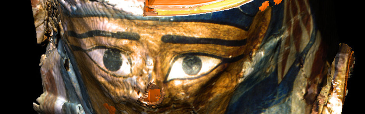
 Since computed tomography uses X-rays, the original colors have naturally been lost. The colors you see on screen are artificial (right). To reflect the mummy’s familiar external appearance, a photograph of the mummy’s mask was projected onto its outer surface (below).
 On the other hand, the mummy is “intelligent”: With the procedure developed in Hamburg, information about objects can be saved in a way which makes it possible to access them in context on screen. For example, an observer can click a certain part of the image to obtain information about the chosen area. This feature is available at the exhibition, but unfortunately not on the World Wide Web.
The method described is very complex in development and execution. Ten million voxels have to be processed to create a new image. For a user not to have to wait to long, a very powerful computer is needed. In this case, a DEC AlphaStation 64-bit UNIX workstation was used. The program used in the exhibition is, therefore, a simplified version, where ready-made image sequences can be called by the the user. This program is also used for the VOXEL-MAN 3D Navigator interactive atlases of anatomy and radiology.
The computer tomographical recordings have kindly been made by the Radiological Clinic of the University Hospital Eppendorf (UKE).
Back to the Virtual Mummy