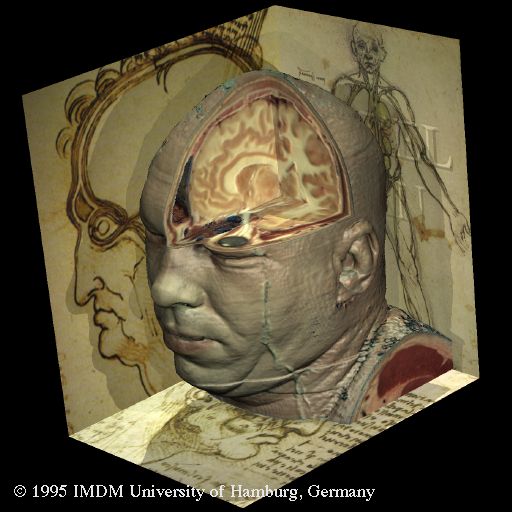
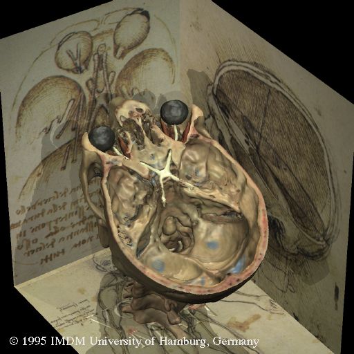
More than 500 years ago, Leonardo da Vinci opened up a new era in the representation and understanding of human anatomy with his famous anatomical drawings. His studies also provided the inspiration for the virtual dissections shown here, created from tomographic images using the VOXEL-MAN 3D visualization system. Both versions show the anatomy in their very own aesthetic.
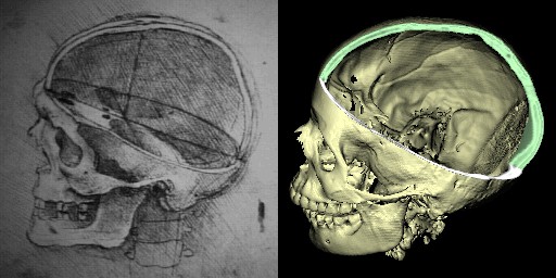
 The skull sectioned (1489, RCIN 919057) | 2300 year old Egyptian mummy reconstructed from CT images (1989)
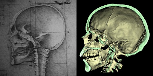
 The cranium (1489, RCIN 919057) | —
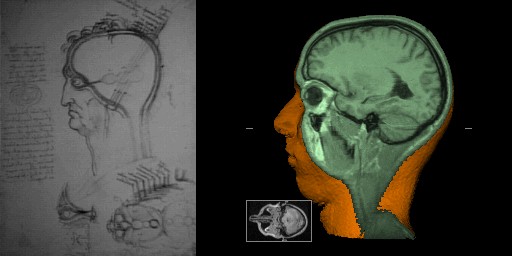 The layers of the scalp, and the cerebral ventricles (ca. 1490-1492, RCIN 912603) | 3D Reconstruction from MRI images (1989) | Please note the small cross-sectional images at the bottom
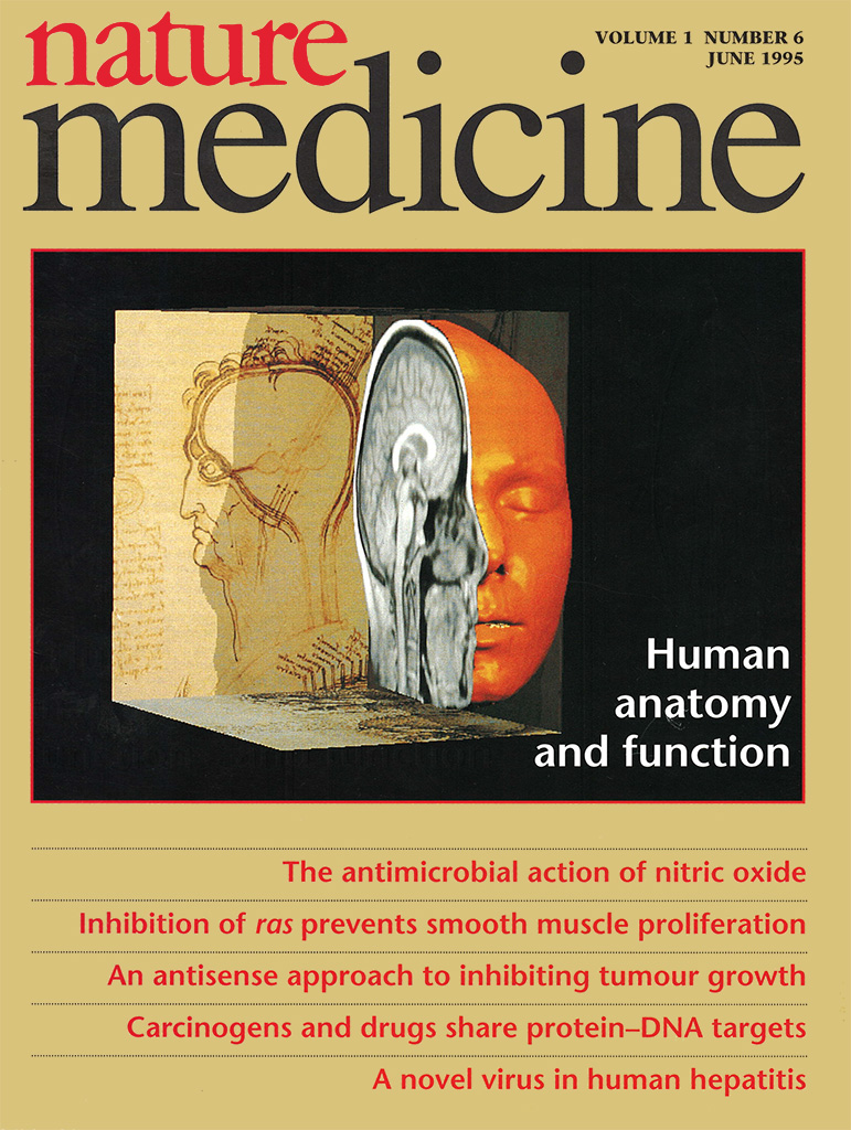 — | 3D Reconstruction from MRI images on the cover of Nature Medicine (1995)
 — | Head of the Visible Human (1995)
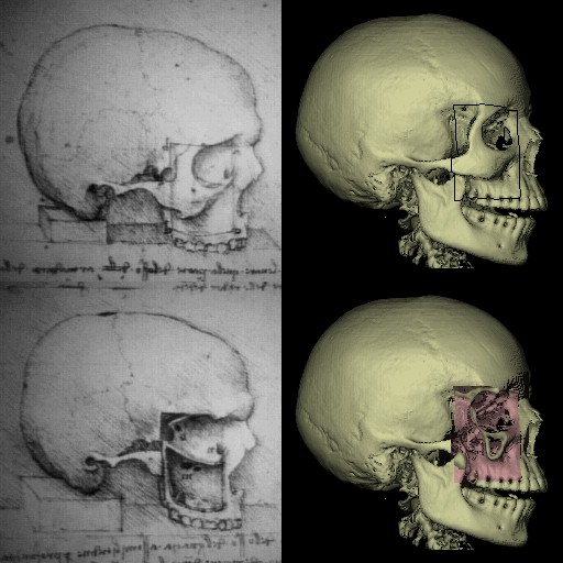
 The cranial nerves (ca. 1508, RCIN 919052) | —
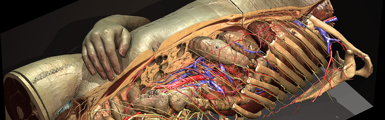
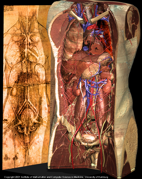 The cardiovascular system and principal organs of a woman (ca. 1509-1510, RCIN 912281) | Torso and internal organs of the Visible Human (2000)
The collection of manuscript sheets by Leonardo da Vinci known as the Codex Windsor is part of the Royal Collection at Windsor Castle. RCIN refers to the inventory number.
References
- Karl Heinz Höhne, Leonardo meets VOXEL-MAN. In Medicine Meets Virtual Reality 17 Course Syllabus: Salon and The Well. Long Beach, CA, 2009, 25.
 Karl Heinz Höhne, Bernhard Pflesser, Andreas Pommert, Martin Riemer, Thomas Schiemann, Rainer Schubert, Ulf Tiede: A new representation of knowledge concerning human anatomy and function. Nature Medicine 1 (6), 1995, 506-511.