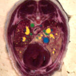
 Inspired by the Visible Human Project, the Faculty of Veterinary Medicine of the Ludwig Maximilian University of Munich launched the Visible Animal Project (VAP). Within the scope of this project, the Visible Dog, a three-dimensional data set of a beagle, was created. The data include colored cryosections (photographic cross-sectional images of the frozen body, right) as well as CT and MRI cross-sectional images. From these data, a 3D model of the dog’s anatomy was created using the VOXEL-MAN visualization system.
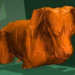
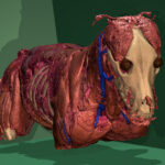
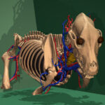
The following image sequence shows the gradual dissection of the Visible Dog from the skin (left) to the superficial musculoskeletal system (center left), the skeleton (center right), and the major blood vessels, bronchi and internal organs (right). The lower parts of the legs are not shown.
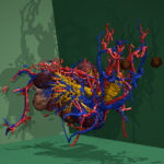
The following illustration shows the internal organs in a lateral view. In shape, relative size and location they differ substantially from the human anatomy. For better visibility, the left ribs were removed with a sagittal cut.
References
- Peter Böttcher: The Visible Animal Project: Virtuelle Realität in der Veterinäranatomie. Dissertation, Tierärztliche Fakultät, Ludwig-Maximilians-Universität München, 2000.
- Peter Böttcher, Johann Maierl, Thomas Schiemann, Cristian Glaser, Renate Weller, Karl Heinz Höhne, Maximilian Reiser, Hans-Georg Liebich: The Visible Animal Project: A three-dimensional, digital database for high quality three-dimensional reconstructions . Veterinary Radiology & Ultrasound 40 (6), 1999, 611-616.
Back to VOXEL-MAN Gallery
