![]()
1994年、アメリカ国立医学図書館のVisible Human Project(ビジブル・ヒューマン・プロジェクト、VHP)は、史上初の人体のトゥルーカラー3次元画像データセットを発表した。
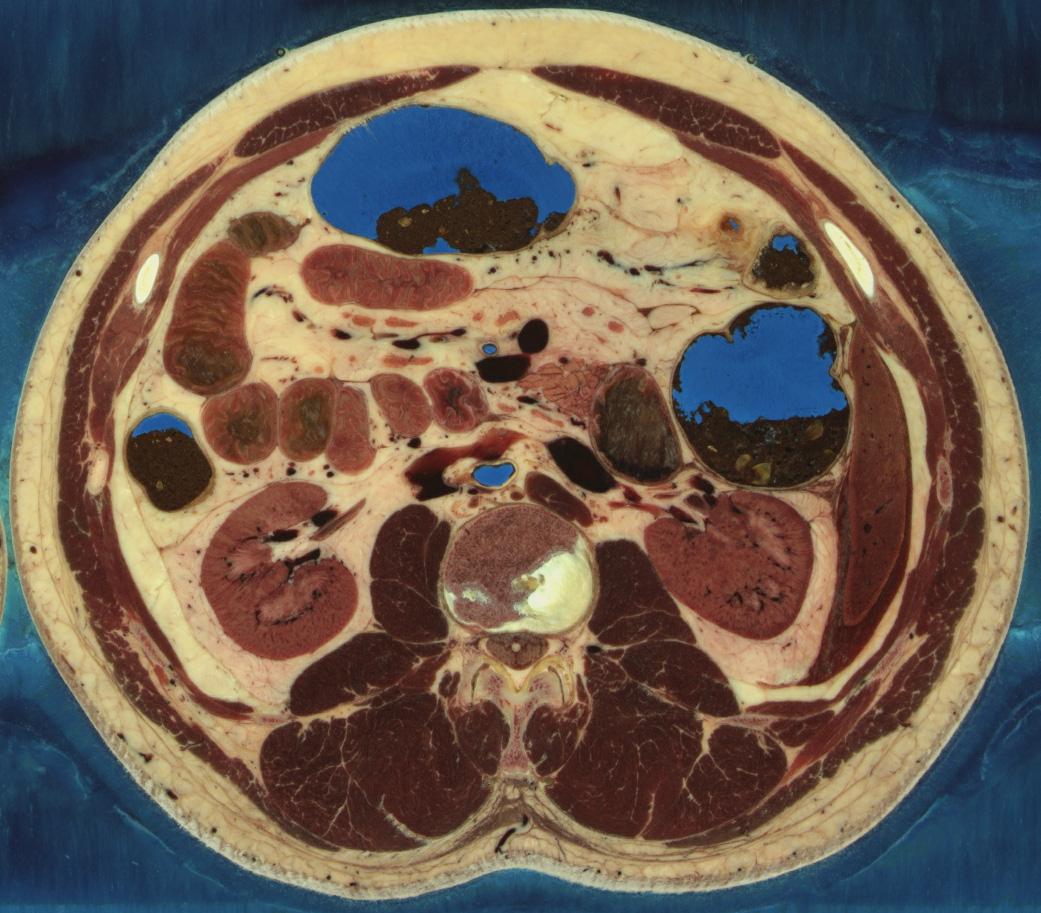
人の遺体を水とゼラチンのブロックに入れて凍らせた。そのブロックから1mmの間隔で薄い層を削り取り、その表面の写真を撮った。こうして、頭からつま先までの人体構造の三次元構造を高解像度と自然な色彩で示す1878枚の横断面画像(凍結切片とも呼ばれる)が作成された。このカラー画像は、過去に取得されたCTやMRI画像によって補完されている。同じ原理に従って、女性の身体のさらに高解像度のデータセットが作成され、画像距離は0.33mmであった。
3Dモデル
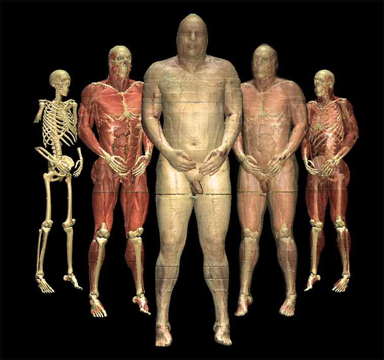
私たちは、可視ヒト男性と可視ヒト女性の断面画像を使って、身体の様々な部位の3Dモデルをこれまでにない詳細さとリアルさで作成しました:
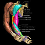 腕と手 (2004)
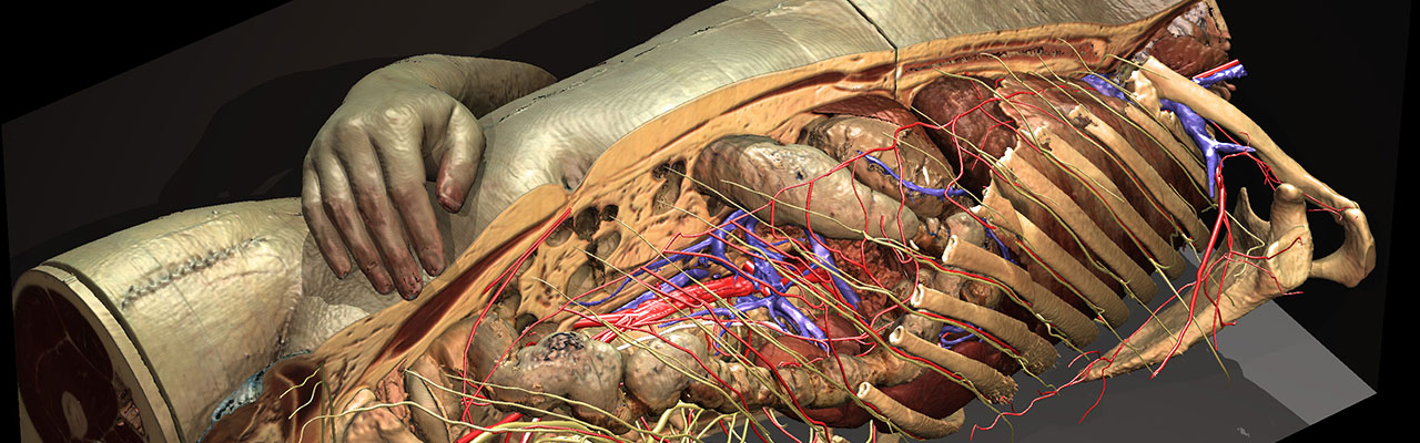
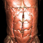 胴体と内臓 (2000)
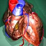 心臓 (1999)
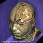 頭部 (1995)
この作業の一部は短編映画で紹介されている。
用途
これらのモデルは、人体解剖学および放射線学の様々なインタラクティブな3Dアトラスの基盤を形成しています。
参考文献
- Thomas Schiemann, Ulf Tiede, Karl Heinz Höhne: Segmentation of the Visible Human for high-quality volume-based visualization. Medical Image Analysis 1 (4), 1997, 263-271.
- Ulf Tiede, Thomas Schiemann, Karl Heinz Höhne: Visualizing the Visible Human. IEEE Computer Graphics and Applications 16 (1), 1996, 7-9.